TIPS was first described in 1969 via balloon dilation between veins.
Indications
Refractory Variceal Bleeding
Refractory Ascites
Hepatorenal syndrome
Preoperative Portal Pressure Reduction
Hepatic Hydrothorax
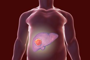
Budd-Chiari syndrome
Contraindications
Severe and progressive liver failure
Polycystic liver disease
Severe encephalopathy
Portal and hepatic vein thrombosis
Pulmonary hypertension
Severe right-heart failure
Hepatopulmonary syndrome
Active infection
Outcomes
TIPS placement success depends on interventional radiologist skill, with data shows over 90% success at three centers.
TIPS placement controls variceal bleeding with a portosystemic gradient under 12 mm Hg.
A gradient under 12 mm Hg lowers variceal bleeding risk and is the target.
Technical failures in portal venous puncture stem from anatomical issues. Ascites reduces significantly within a month in 50-90% of cases.
Late stenosis and occlusion are linked to pseudointimal or intimal hyperplasia in stents.
Equipment required
Good ultrasound machine with linear-array probe
Fluoroscopy
Basic angiography set
Sheath and curved catheter
Guide wires
Pressure transducer
Angioplasty balloons
Patient Preparation:
General anesthesia is common for pediatric and often preferred for adults.
Midazolam and fentanyl citrate are effective for sedation. Local anesthesia achieved with 5 mL lidocaine 1% at jugular.
Impaired renal function requires pre-hydration and lower osmolality non-ionic contrast agents.
Pre-procedure imaging ensures portal and hepatic vein patency while ruling out tumors in the area.
Large-volume ascites can be drained prior to the procedure for advanced techniques like percutaneous guide wire placement.
Informed Consent:
Explain the procedure’s risks and potential complications clearly to the patient.
Patient Positioning:
The patient should be positioned in supine position with their neck turned away from the side of vein puncture.
Technique
Step 1: Venous Access and Catheter Placement
A sheath is inserted into the right internal jugular vein. Then a guiding catheter is advanced into the hepatic vein.
Step 2: Portal Vein Puncture
With help of fluoroscopy and ultrasound, a TIPS needle system is used to puncture the portal vein from the hepatic vein.
Step 3: Tract Dilation
Guidewire is advanced into the portal vein very carefully for stability.
The tract is dilated using a balloon catheter to widen the connection.
Step 4: Stent Placement
A self-expandable stent should be placed to maintain the shunt between the hepatic and portal veins.
Step 5: Hemodynamic Assessment
Finally portal pressure gradient is measured before and after TIPS creation.
TIPS was first described in 1969 via balloon dilation between veins.
Refractory Variceal Bleeding
Refractory Ascites
Hepatorenal syndrome
Preoperative Portal Pressure Reduction
Hepatic Hydrothorax
Budd-Chiari syndrome
Severe and progressive liver failure
Polycystic liver disease
Severe encephalopathy
Portal and hepatic vein thrombosis
Pulmonary hypertension
Severe right-heart failure
Hepatopulmonary syndrome
Active infection
TIPS placement success depends on interventional radiologist skill, with data shows over 90% success at three centers.
TIPS placement controls variceal bleeding with a portosystemic gradient under 12 mm Hg.
A gradient under 12 mm Hg lowers variceal bleeding risk and is the target.
Technical failures in portal venous puncture stem from anatomical issues. Ascites reduces significantly within a month in 50-90% of cases.
Late stenosis and occlusion are linked to pseudointimal or intimal hyperplasia in stents.
Good ultrasound machine with linear-array probe
Fluoroscopy
Basic angiography set
Sheath and curved catheter
Guide wires
Pressure transducer
Angioplasty balloons
Patient Preparation:
General anesthesia is common for pediatric and often preferred for adults.
Midazolam and fentanyl citrate are effective for sedation. Local anesthesia achieved with 5 mL lidocaine 1% at jugular.
Impaired renal function requires pre-hydration and lower osmolality non-ionic contrast agents.
Pre-procedure imaging ensures portal and hepatic vein patency while ruling out tumors in the area.
Large-volume ascites can be drained prior to the procedure for advanced techniques like percutaneous guide wire placement.
Informed Consent:
Explain the procedure’s risks and potential complications clearly to the patient.
Patient Positioning:
The patient should be positioned in supine position with their neck turned away from the side of vein puncture.
Step 1: Venous Access and Catheter Placement
A sheath is inserted into the right internal jugular vein. Then a guiding catheter is advanced into the hepatic vein.
Step 2: Portal Vein Puncture
With help of fluoroscopy and ultrasound, a TIPS needle system is used to puncture the portal vein from the hepatic vein.
Step 3: Tract Dilation
Guidewire is advanced into the portal vein very carefully for stability.
The tract is dilated using a balloon catheter to widen the connection.
Step 4: Stent Placement
A self-expandable stent should be placed to maintain the shunt between the hepatic and portal veins.
Step 5: Hemodynamic Assessment
Finally portal pressure gradient is measured before and after TIPS creation.
Both our subscription plans include Free CME/CPD AMA PRA Category 1 credits.
Digital Certificate PDF
On course completion, you will receive a full-sized presentation quality digital certificate.
medtigo Simulation
A dynamic medical simulation platform designed to train healthcare professionals and students to effectively run code situations through an immersive hands-on experience in a live, interactive 3D environment.
medtigo Points
medtigo points is our unique point redemption system created to award users for interacting on our site. These points can be redeemed for special discounts on the medtigo marketplace as well as towards the membership cost itself.
Community Forum post/reply = 5 points
*Redemption of points can occur only through the medtigo marketplace, courses, or simulation system. Money will not be credited to your bank account. 10 points = $1.
All Your Certificates in One Place
When you have your licenses, certificates and CMEs in one place, it's easier to track your career growth. You can easily share these with hospitals as well, using your medtigo app.